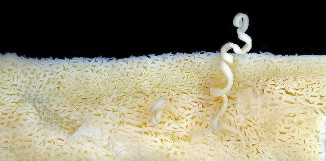
Tapeworm Parasite in Mustelus canis panorama attached to intestine

$19.00$400.00

Cestoda (Cestoidea) is a class of parasitic worms of the flatworm(Platyhelminthes) phylum. They are informally referred to as cestodes. The best-known species are commonly called tapeworms. All cestodes are parasitic and their life histories vary, but typically they live in the digestive tracts of vertebrates as adults, and often in the bodies of other species of animals as juveniles. Over a thousand species have been described, and all vertebrate species may be parasitised by at least one species of tapeworm.

Tapeworm Theme: Research by Jimmy Bernot and Dr. Janine Caira

Researchers in Dr. Janine Caira’s lab at UConn study tapeworms: the ribbon-like parasites found in the intestine of all major groups of vertebrates. The Caira lab primarily studies tapeworms from sharks, skates, and stingrays, which host a diverse assortment of tapeworms. We are interested in how these tapeworms are related, how they evolve, and the dynamic interactions they have with their hosts over time.
It may seem rare or even shocking to discover a new species, but in the Caira lab numerous new species of tapeworms are discovered and described just about every year! In fact, the tapeworms photographed here by Mark Smith were discovered by the Caira lab in our very own back yard — Long Island Sound. This species is found in the intestine of a common shark, Mustelus canis, often called the smoothdog fish, which may be familiar to recreational fisherman in the Northeastern U.S. In fact, if you have ever caught a smoothdog fish, it is likely that the tapeworm species pictured here was inside the shark you caught, just waiting to be discovered!

One of the main goals of my Master’s thesis was to understand how this species remains attached to the intestine of its shark host. This is of the utmost importance to these worms because they cannot survive outside of the their host’s intestine. These photographs where taken to better understand the way these tapeworms maintain this intimate relationship.

In these close-up images of its scolex (think of it as the tapeworm equivalent of a head, yet strangely lacking eyes, ears, a nose and a mouth) you can see many of the specialized structures like hooks, muscular flaps, and suckers that enable this worm to remain securely anchored to its host. If you look along its ribbon-like body, you can see serrated edges characteristic of this tapeworm and its relatives.

In these photographs you can see tapeworms attached to the intestine of a shark. These photographs have helped us understand that the serrated margins of these worms help secure the worm to its host by locking into small surface features of the intestine like dovetail joints — something never before shown — demonstrating the functionality of these previously mysterious structures.

Together, these photographs help us better understand the way these worms live their bizarre lives in the dark, complex, and shifting environment of a shark intestine.

Description

Commercial Use

Images may be licensed for commercial use via the “add-to-cart” button on each of the image pages.

Non-Profit, Educational & Academic Use

All of our images are “Creative Commons” for non-profit, educational and academic use. Fill out this form and please provide a visible photo credit to Macroscopic Solutions with a link to www.macroscopicsolutions.com when using our images for this purpose.

Additional information

Size

150×150 ($19), 240×160 ($29), 640×427 ($49), 2048×1365 ($199), Original ($400)

Reviews

There are no reviews yet.

Be the first to review “Tapeworm Parasite in Mustelus canis panorama attached to intestine”