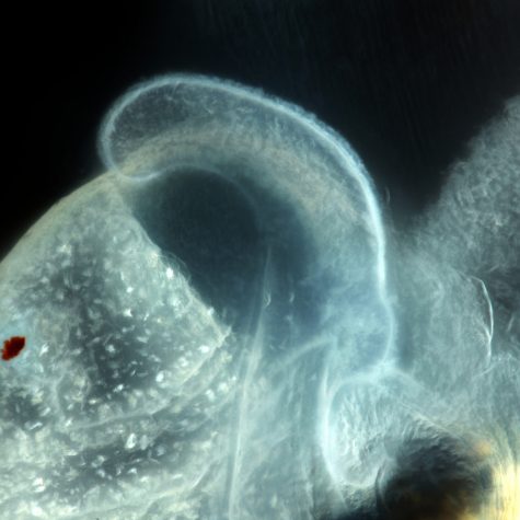
Fin Structure of a Cleared and Stained Fish, Hudson River Valley

$19.00$400.00

Staining is an auxiliary technique used in microscopy to enhance contrast in the microscopic image. Stains and dyes are frequently used in biology and medicine to highlight structures in biological tissues for viewing, often with the aid of different microscopes. Stains may be used to define and examine bulk tissues (highlighting, for example, muscle fibers or connective tissue), cell populations (classifying different blood cells, for instance), or organelles within individual cells.

Description

Commercial Use

Images may be licensed for commercial use via the “add-to-cart” button on each of the image pages.

Non-Profit, Educational & Academic Use

All of our images are “Creative Commons” for non-profit, educational and academic use. Fill out this form and please provide a visible photo credit to Macroscopic Solutions with a link to www.macroscopicsolutions.com when using our images for this purpose.

Additional information

Size

150×150 ($19), 240×160 ($29), 640×427 ($49), 2048×1365 ($199), Original ($400)

Reviews

There are no reviews yet.

Be the first to review “Fin Structure of a Cleared and Stained Fish, Hudson River Valley”