Showing 1–16 of 62 resultsSorted by price: high to low
Select options
This product has multiple variants. The options may be chosen on the product page
$19.00–$400.00Price range: $19.00 through $400.00
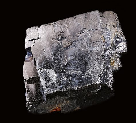
Galena
Galena is the main ore of lead, used since ancient times. Because of its somewhat low melting point, it was easy to liberate by smelting. It typically forms in low-temperature sedimentary deposits.
In some deposits galena contains about 1–2% silver, a byproduct that far outweighs the main lead ore in revenue. Galena deposits often also contain significant amounts of silver as included silver sulfide mineral phases or as limited solid solution within the galena structure. These argentiferous galenas have long been the most important ore of silver.[citation needed]
Cubic galena with calcite from Jasper County, Missouri, USA; 5.1 cm × 3.2 cm × 2.8 cm (2.0 in × 1.3 in × 1.1 in)
Galena also was a major mineral of the zinc-lead mines of the tri-state district around Joplin in southwestern Missouri and the adjoining areas of Kansas and Oklahoma. Galena is also an important ore mineral in the silver mining regions of Colorado, Idaho, Utah and Montana. Of the latter, the Coeur d’Alene district of northern Idaho was most prominent.
Derbyshire in the UK was one of the main areas where galena was mined.
The largest documented crystal of galena is composite cubo-octahedra from the Great Laxey Mine, Isle of Man, measuring 25 cm × 25 cm × 25 cm (10 in × 10 in × 10 in).
Select options
This product has multiple variants. The options may be chosen on the product page
$19.00–$400.00Price range: $19.00 through $400.00
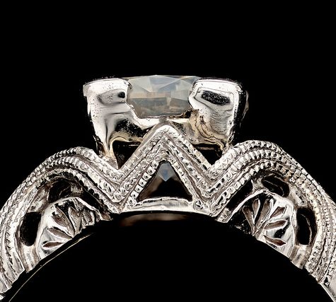
Fancy White Diamond 1.08 Ct in Victorian Setting
A chemically pure and structurally perfect diamond is perfectly transparent with no hue, or color. However, in reality almost no gem-sized natural diamonds are absolutely perfect. The color of a diamond may be affected by chemical impuritiesand/or structural defects in the crystal lattice. Depending on the hue and intensity of a diamond’s coloration, a diamond’s color can either detract from or enhance its value. For example, most white diamonds are discounted in price when more yellow hue is detectable, while intense pink diamonds or blue diamonds (such as the Hope Diamond) can be dramatically more valuable. Of all colored diamonds, red diamonds are the rarest. The Aurora Pyramid of Hope displays a spectacular array of naturally colored diamonds, including red diamonds.
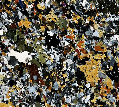
When placed between two polarizing filters set at right angles to each other, the optical properties of the minerals in the thin section alter the colour and intensity of the light as seen by the viewer. As different minerals have different optical properties, most rock forming minerals can be easily identified. Plagioclase for example can be seen in the photo on the right as a clear mineral with multiple parallel twinning planes. The large blue-green minerals are clinopyroxene with some exsolution of orthopyroxene.
Thin sections are prepared in order to investigate the optical properties of the minerals in the rock. This work is a part of petrology and helps to reveal the origin and evolution of the parent rock.
A photograph of a rock in thin section is often referred to as a photomicrograph.
Select options
This product has multiple variants. The options may be chosen on the product page
$19.00–$400.00Price range: $19.00 through $400.00
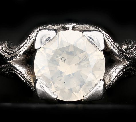
Fancy White Diamond 1.08 Ct in Victorian Setting
A chemically pure and structurally perfect diamond is perfectly transparent with no hue, or color. However, in reality almost no gem-sized natural diamonds are absolutely perfect. The color of a diamond may be affected by chemical impuritiesand/or structural defects in the crystal lattice. Depending on the hue and intensity of a diamond’s coloration, a diamond’s color can either detract from or enhance its value. For example, most white diamonds are discounted in price when more yellow hue is detectable, while intense pink diamonds or blue diamonds (such as the Hope Diamond) can be dramatically more valuable. Of all colored diamonds, red diamonds are the rarest. The Aurora Pyramid of Hope displays a spectacular array of naturally colored diamonds, including red diamonds.
When placed between two polarizing filters set at right angles to each other, the optical properties of the minerals in the thin section alter the colour and intensity of the light as seen by the viewer. As different minerals have different optical properties, most rock forming minerals can be easily identified. Plagioclase for example can be seen in the photo on the right as a clear mineral with multiple parallel twinning planes. The large blue-green minerals are clinopyroxene with some exsolution of orthopyroxene.
Thin sections are prepared in order to investigate the optical properties of the minerals in the rock. This work is a part of petrology and helps to reveal the origin and evolution of the parent rock.
A photograph of a rock in thin section is often referred to as a photomicrograph.
Select options
This product has multiple variants. The options may be chosen on the product page
$19.00–$400.00Price range: $19.00 through $400.00
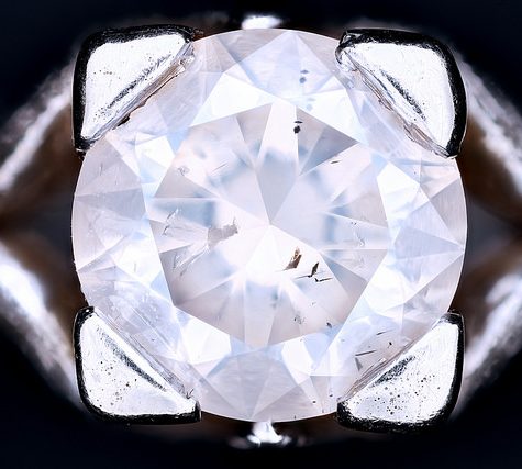
Fancy White Diamond 1.08 Ct in Victorian Setting
A chemically pure and structurally perfect diamond is perfectly transparent with no hue, or color. However, in reality almost no gem-sized natural diamonds are absolutely perfect. The color of a diamond may be affected by chemical impuritiesand/or structural defects in the crystal lattice. Depending on the hue and intensity of a diamond’s coloration, a diamond’s color can either detract from or enhance its value. For example, most white diamonds are discounted in price when more yellow hue is detectable, while intense pink diamonds or blue diamonds (such as the Hope Diamond) can be dramatically more valuable. Of all colored diamonds, red diamonds are the rarest. The Aurora Pyramid of Hope displays a spectacular array of naturally colored diamonds, including red diamonds.
When placed between two polarizing filters set at right angles to each other, the optical properties of the minerals in the thin section alter the colour and intensity of the light as seen by the viewer. As different minerals have different optical properties, most rock forming minerals can be easily identified. Plagioclase for example can be seen in the photo on the right as a clear mineral with multiple parallel twinning planes. The large blue-green minerals are clinopyroxene with some exsolution of orthopyroxene.
Thin sections are prepared in order to investigate the optical properties of the minerals in the rock. This work is a part of petrology and helps to reveal the origin and evolution of the parent rock.
A photograph of a rock in thin section is often referred to as a photomicrograph.
Select options
This product has multiple variants. The options may be chosen on the product page
$19.00–$400.00Price range: $19.00 through $400.00
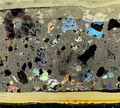
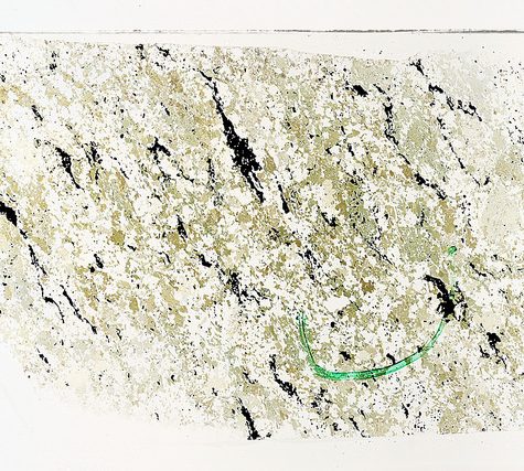
Reunion Island, 22,189, Old Glue disrupted optical properties 1x
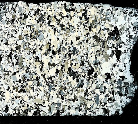
In optical mineralogy and petrography, a thin section (or petrographic thin section) is a laboratory preparation of a rock, mineral, soil, pottery, bones, or even metal sample for use with a polarizingpetrographic microscope, electron microscope and electron microprobe. A thin sliver of rock is cut from the sample with a diamond saw and ground optically flat. It is then mounted on a glass slide and then ground smooth using progressively finer abrasive grit until the sample is only 30 μm thick. The method involved using the Michel-Lévy interference colour chart. Typically quartz is used as the gauge to determine thickness as it is one of the most abundant minerals.
When placed between two polarizing filters set at right angles to each other, the optical properties of the minerals in the thin section alter the colour and intensity of the light as seen by the viewer. As different minerals have different optical properties, most rock forming minerals can be easily identified. Plagioclase for example can be seen in the photo on the right as a clear mineral with multiple parallel twinning planes. The large blue-green minerals are clinopyroxene with some exsolution of orthopyroxene.
Thin sections are prepared in order to investigate the optical properties of the minerals in the rock. This work is a part of petrology and helps to reveal the origin and evolution of the parent rock.
A photograph of a rock in thin section is often referred to as a photomicrograph.
Select options
This product has multiple variants. The options may be chosen on the product page
$19.00–$400.00Price range: $19.00 through $400.00
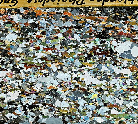
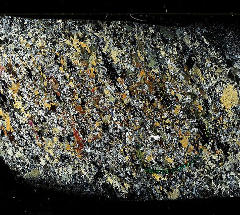
Thin Section, 5x Polarized
In optical mineralogy and petrography, a thin section (or petrographic thin section) is a laboratory preparation of a rock, mineral, soil, pottery, bones, or even metal sample for use with a polarizingpetrographic microscope, electron microscope and electron microprobe. A thin sliver of rock is cut from the sample with a diamond saw and ground optically flat. It is then mounted on a glass slide and then ground smooth using progressively finer abrasive grit until the sample is only 30 μm thick. The method involved using the Michel-Lévy interference colour chart. Typically quartz is used as the gauge to determine thickness as it is one of the most abundant minerals.
When placed between two polarizing filters set at right angles to each other, the optical properties of the minerals in the thin section alter the colour and intensity of the light as seen by the viewer. As different minerals have different optical properties, most rock forming minerals can be easily identified. Plagioclase for example can be seen in the photo on the right as a clear mineral with multiple parallel twinning planes. The large blue-green minerals are clinopyroxene with some exsolution of orthopyroxene.
Thin sections are prepared in order to investigate the optical properties of the minerals in the rock. This work is a part of petrology and helps to reveal the origin and evolution of the parent rock.
A photograph of a rock in thin section is often referred to as a photomicrograph.
Select options
This product has multiple variants. The options may be chosen on the product page
$19.00–$400.00Price range: $19.00 through $400.00
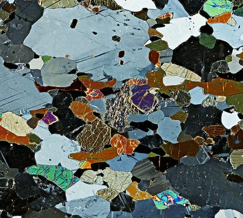
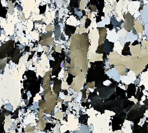
Thin Section, 1x
In optical mineralogy and petrography, a thin section (or petrographic thin section) is a laboratory preparation of a rock, mineral, soil, pottery, bones, or even metal sample for use with a polarizingpetrographic microscope, electron microscope and electron microprobe. A thin sliver of rock is cut from the sample with a diamond saw and ground optically flat. It is then mounted on a glass slide and then ground smooth using progressively finer abrasive grit until the sample is only 30 μm thick. The method involved using the Michel-Lévy interference colour chart. Typically quartz is used as the gauge to determine thickness as it is one of the most abundant minerals.
When placed between two polarizing filters set at right angles to each other, the optical properties of the minerals in the thin section alter the colour and intensity of the light as seen by the viewer. As different minerals have different optical properties, most rock forming minerals can be easily identified. Plagioclase for example can be seen in the photo on the right as a clear mineral with multiple parallel twinning planes. The large blue-green minerals are clinopyroxene with some exsolution of orthopyroxene.
Thin sections are prepared in order to investigate the optical properties of the minerals in the rock. This work is a part of petrology and helps to reveal the origin and evolution of the parent rock.
A photograph of a rock in thin section is often referred to as a photomicrograph.
Select options
This product has multiple variants. The options may be chosen on the product page
$19.00–$400.00Price range: $19.00 through $400.00
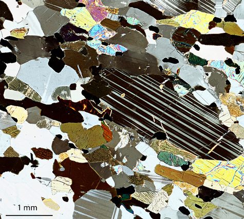
Thin Section, 1x Polarized
In optical mineralogy and petrography, a thin section (or petrographic thin section) is a laboratory preparation of a rock, mineral, soil, pottery, bones, or even metal sample for use with a polarizingpetrographic microscope, electron microscope and electron microprobe. A thin sliver of rock is cut from the sample with a diamond saw and ground optically flat. It is then mounted on a glass slide and then ground smooth using progressively finer abrasive grit until the sample is only 30 μm thick. The method involved using the Michel-Lévy interference colour chart. Typically quartz is used as the gauge to determine thickness as it is one of the most abundant minerals.
When placed between two polarizing filters set at right angles to each other, the optical properties of the minerals in the thin section alter the colour and intensity of the light as seen by the viewer. As different minerals have different optical properties, most rock forming minerals can be easily identified. Plagioclase for example can be seen in the photo on the right as a clear mineral with multiple parallel twinning planes. The large blue-green minerals are clinopyroxene with some exsolution of orthopyroxene.
Thin sections are prepared in order to investigate the optical properties of the minerals in the rock. This work is a part of petrology and helps to reveal the origin and evolution of the parent rock.
A photograph of a rock in thin section is often referred to as a photomicrograph.
Select options
This product has multiple variants. The options may be chosen on the product page
$19.00–$400.00Price range: $19.00 through $400.00
Thin Section, 5x Polarized
In optical mineralogy and petrography, a thin section (or petrographic thin section) is a laboratory preparation of a rock, mineral, soil, pottery, bones, or even metal sample for use with a polarizingpetrographic microscope, electron microscope and electron microprobe. A thin sliver of rock is cut from the sample with a diamond saw and ground optically flat. It is then mounted on a glass slide and then ground smooth using progressively finer abrasive grit until the sample is only 30 μm thick. The method involved using the Michel-Lévy interference colour chart. Typically quartz is used as the gauge to determine thickness as it is one of the most abundant minerals.
When placed between two polarizing filters set at right angles to each other, the optical properties of the minerals in the thin section alter the colour and intensity of the light as seen by the viewer. As different minerals have different optical properties, most rock forming minerals can be easily identified. Plagioclase for example can be seen in the photo on the right as a clear mineral with multiple parallel twinning planes. The large blue-green minerals are clinopyroxene with some exsolution of orthopyroxene.
Thin sections are prepared in order to investigate the optical properties of the minerals in the rock. This work is a part of petrology and helps to reveal the origin and evolution of the parent rock.
A photograph of a rock in thin section is often referred to as a photomicrograph.
Select options
This product has multiple variants. The options may be chosen on the product page
$19.00–$400.00Price range: $19.00 through $400.00
Thin Section, 1x Polarized
In optical mineralogy and petrography, a thin section (or petrographic thin section) is a laboratory preparation of a rock, mineral, soil, pottery, bones, or even metal sample for use with a polarizingpetrographic microscope, electron microscope and electron microprobe. A thin sliver of rock is cut from the sample with a diamond saw and ground optically flat. It is then mounted on a glass slide and then ground smooth using progressively finer abrasive grit until the sample is only 30 μm thick. The method involved using the Michel-Lévy interference colour chart. Typically quartz is used as the gauge to determine thickness as it is one of the most abundant minerals.
When placed between two polarizing filters set at right angles to each other, the optical properties of the minerals in the thin section alter the colour and intensity of the light as seen by the viewer. As different minerals have different optical properties, most rock forming minerals can be easily identified. Plagioclase for example can be seen in the photo on the right as a clear mineral with multiple parallel twinning planes. The large blue-green minerals are clinopyroxene with some exsolution of orthopyroxene.
Thin sections are prepared in order to investigate the optical properties of the minerals in the rock. This work is a part of petrology and helps to reveal the origin and evolution of the parent rock.
A photograph of a rock in thin section is often referred to as a photomicrograph.