Macropod PRO 3D R5 Educational (Excludes 7.5-100x Objectives)

Macropod Petrographic
The Macropod PETROGRAPHIC is a rigid photomacrography system that allows slides/thin sections to be imaged and analyzed in plain light and cross polarized light.

The Macropod PETROGRAPHIC is a rigid photomacrography system that allows slides/thin sections to be imaged and analyzed in plain light and cross polarized light.
Basic Support is provided to all of Macroscopic Solutions customers for a limited period of time. These requests are handled over the phone, by email or by web video. They are available for 30 days after the date of purchase, there is a fee associated with assistance related to training beyond the 30 day time period. For those seeking additional, on-site and hands-on training and support, please consider purchasing the extended, premium or international support package.
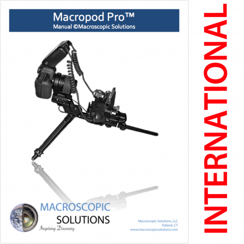
International Support is available upon request, please contact support@macroscopicsolutions.com for more information. A representative will respond within 1 hour during regular business hours (US EST or EDT).
International support includes all insured delivery fees and a 3 day training program for the client. The client can invite affiliated faculty, students and other associates for one-on-one training sessions. These trains sessions allow a wide range of Macropod users to become specialists at imaging and 3D modeling the specimens they regularly work with.
Macroscopic Solutions will send a technician approximately 1 month after the date of delivery. This allows the client to gather some experience prior to the on-site training, giving the client further insight into the types of questions they should be asking.
Macroscopic Solutions technicians maintain close contact after on-site training occurs to ensure proper handling and use of the system.
Basic Support is provided to all of Macroscopic Solutions customers for a limited period of time. These requests are handled over the phone, by email or by web video. They are available for 30 days after the date of purchase, there is a fee associated with assistance related to training beyond the 30 day time period. For those seeking additional, on-site and hands-on training and support, please consider purchasing the extended or premium support package for you.
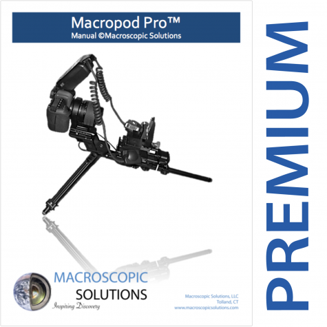
Premium Support is provided to customers who would like to train multiple members of their research group over an extended period of time. A trained professional from Macroscopic Solutions will visit the customers location subsequent to delivery in order to describe the capabilities and train all colleagues wishing to use Macroscopic Solutions systems. This is one of two provided on site visitations included with Premium Support. Support by phone, email and web video is extended from a 30 day period to a 3 year period for any customers acquiring immediate assistance. A second onsite visitation is also included with the Premium Support package. This is helpful when components of Macroscopic Solutions systems are upgraded throughout time or the customer has an influx of new colleagues, employees, students, etc. to train that were not present during the initial training and support session.
For any researcher, it is important to fully understand how the Macropod works best for you. With your purchase, we offer three levels of support to meet your personal imaging needs: Basic, Extended, or Premium.
Extended Bundle:
Comes with personal delivery, set-up and demonstration; and online consultations and support for 1 year.
Premium Bundle:
Comes with personal delivery, set-up and demonstration; secondary visitation upon request; and online consultations and support for 3 years.
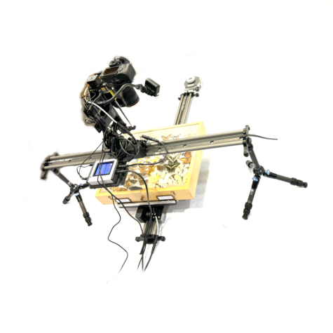
Bring industrial precision to scientific imaging with the Macropod Core 3rd Axes Conversion — an advanced upgrade enabling large-format panoramic imaging, 3-axis automation, and precision focus stacking. This system is engineered for clients requiring high-resolution documentation of core boxes, entomology drawers, tree cores, soil boxes, and other elongated specimens with substantial length and width.
Full 3-Axis Automation: Adds a third linear rail to your Macropod Core system for complete X-Y-Z motion control, enabling seamless panoramic captures and automated focus stacking across large subjects.
Panoramic Imaging Workflow: Perfect for digitizing entire drawers or trays, the 3rd axis ensures complete coverage without manual repositioning.
Unmatched Precision: Each axis is guided by carbon-fiber rails and tension-cable alignment, ensuring micrometer-level accuracy across large surfaces.
Broad Compatibility: Designed for core box imaging, whole-drawer insect digitization, geological core photography, and botanical tray scanning.
High-Resolution Outputs: Produces photogrammetric datasets and stitched mosaics suitable for measurement, AI training, or 3D modeling.
Open Workflow: Integrates with software like Agisoft Metashape, Zerene Stacker, and Helicon Focus for photogrammetry and image stacking.
The complete playlist can be viewed on our YouTube Channel here: https://www.youtube.com/watch?v=Whoa4jDUYo8&list=PLcKIGvtblfut6S-o7jE01siFfMwwFHQsO
Basic Support is provided to all of Macroscopic Solutions customers for a limited period of time. These requests are handled over the phone, by email or by web video. They are available for 30 days after the date of purchase, there is a fee associated with assistance related to training beyond the 30 day time period. For those seeking additional, on-site and hands-on training and support, please consider purchasing the extended or premium support package for you.
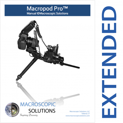
Extended Support is provided to customers who would like to train multiple members of their research group over an extended period of time. A trained professional from Macroscopic Solutions will visit the customers location subsequent to delivery in order to describe the capabilities and train all colleagues wishing to use Macroscopic Solutions systems. Support by phone, email and web video is extended from a 30 day period to a 1 year period for any customers acquiring immediate assistance.
For any researcher, it is important to fully understand how the Macropod works best for you. With your purchase, we offer three levels of support to meet your personal imaging needs: Basic, Extended, or Premium.
Extended Bundle:
Comes with personal delivery, set-up and demonstration; and online consultations and support for 1 year.
Premium Bundle:
Comes with personal delivery, set-up and demonstration; secondary visitation upon request; and online consultations and support for 3 years.
If a hardware component of the Macropod product line is in need of repair and out of warranty, please purchase a repair request and send your part to:
Macroscopic Solutions
ATTN: Repairs
222 Pitkin St
East Hartford, CT 06108
The total cost covers component repair, testing, cleaning and return postage. If additional parts or repairs are necessary, we will inform you of any balance due prior to starting the work.
Description of Problem/Symptoms
Camera shows Error 20 or Error 30. Camera needs Mirror Box Ass’y. Camera will be checked, cleaned and repaired to good working order and functional test. If additional parts are needed, an updated balance will be sent subsequent to payment.
Send with the following accessories: Body Cap, Eyecup, No Battery, No Media Card
|
Description |
Gross Price |
Net Price |
|
Labor |
$398.00 |
$398.00 |
|
Parts & Return Shipping |
$182.43 |
$182.43 |
|
Total Charge |
$580.43 |
Thank you for choosing Macroscopic Solutions!
If a hardware component of the Macropod product line is in need of repair and out of warranty, please purchase a repair request and send your part to:
Macroscopic Solutions
ATTN: Repairs
1 Technology Dr
Tolland CT 06084
The total cost covers component repair, testing, cleaning and return postage.
Thank you for choosing Macroscopic Solutions!
The Nyholm lab studies beneficial host-microbe interactions between the Hawaiian bobtail squid, Euprymna scolopes, and the bioluminescent bacterium, Vibrio fischeri. Hawaiian bobtail squid are nocturnal predators, remaining buried under the sand during the day and coming out to hunt for shrimp at night neat coral reefs. The squid have a light organ on their underside that houses a colony of glowing bacteria (V. fischeri). The squid uses this bacterial bioluminescence in a form of camouflage called counter-illumination, masking it’s silhouette by matching moonlight and starlight; thus hiding from predators swimming below. The light organ is attached to the ink sac and it can use this ink like a type of shutter to control the amount of light. This likely helps the squid adjust to variable light conditions, for example cloudy nights or a full vs. new moon. In this image of a juvenile squid, you can clearly see the bi-lobed light organ and ink sac in the center of the squid’s mantle cavity.
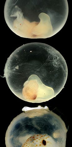
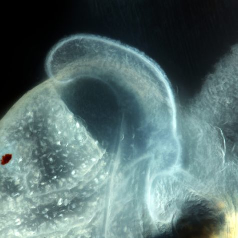
The Hawaiian bobtail squid lay their eggs in clutches on the sea floor, where they take approximately three weeks to develop. This series of macropod images allows us to see the developing squid and monitor embryogenesis. Once the squid hatch, V. fischeri from seawater colonize the light organ within hours. This macropod image allows us to see a close-up view of the ciliated appendage-like structure found on the surface of the juvenile squid’s light organ. Once the squid hatches, the cilia assist in bringing V. fischeri in the seawater to pores at the base of the light organ. These pores lead to inner crypts, where only V. fischeri can enter and colonize. V. fischeri is a relatively rare member of the seawater bacterial community, making up less than 0.1%. The Nyholm lab is trying to understand how the squid’s immune system can differentiate between the symbiont and all the other different kinds of bacteria in seawater.
While the light organ of the squid exemplifies a highly specific beneficial relationship between bacteria and host to provide camouflage at night, this organ is only found in some squid species. All squid, however, are capable of another type of camouflage, cryptic coloration. Squid skin contains special pigmented cells called chromatophores that can change the overall color of the squid in seconds. Each chromatophore contains pigment granules surrounded by nerve and muscle fibers. When these muscles are contracted, the pigment sac expands, creating a larger surface area of color. When the muscles relax, the pigment sac can shrink to a small dot, 15 times smaller than their expanded size, hiding the color. In these macropod images you can see relaxed chromatophores on the mantle and contracted chromatophores around the eyes. The macropod images allow us to see these pigment cells in great detail.
If a hardware component of the Macropod product line is in need of repair and out of warranty, please purchase a repair request and send your part to:
The total cost covers component repair, testing, cleaning and return postage.
Thank you for choosing Macroscopic Solutions!
The Nyholm lab studies beneficial host-microbe interactions between the Hawaiian bobtail squid, Euprymna scolopes, and the bioluminescent bacterium, Vibrio fischeri. Hawaiian bobtail squid are nocturnal predators, remaining buried under the sand during the day and coming out to hunt for shrimp at night neat coral reefs. The squid have a light organ on their underside that houses a colony of glowing bacteria (V. fischeri). The squid uses this bacterial bioluminescence in a form of camouflage called counter-illumination, masking it’s silhouette by matching moonlight and starlight; thus hiding from predators swimming below. The light organ is attached to the ink sac and it can use this ink like a type of shutter to control the amount of light. This likely helps the squid adjust to variable light conditions, for example cloudy nights or a full vs. new moon. In this image of a juvenile squid, you can clearly see the bi-lobed light organ and ink sac in the center of the squid’s mantle cavity.
The Hawaiian bobtail squid lay their eggs in clutches on the sea floor, where they take approximately three weeks to develop. This series of macropod images allows us to see the developing squid and monitor embryogenesis. Once the squid hatch, V. fischeri from seawater colonize the light organ within hours. This macropod image allows us to see a close-up view of the ciliated appendage-like structure found on the surface of the juvenile squid’s light organ. Once the squid hatches, the cilia assist in bringing V. fischeri in the seawater to pores at the base of the light organ. These pores lead to inner crypts, where only V. fischeri can enter and colonize. V. fischeri is a relatively rare member of the seawater bacterial community, making up less than 0.1%. The Nyholm lab is trying to understand how the squid’s immune system can differentiate between the symbiont and all the other different kinds of bacteria in seawater.
While the light organ of the squid exemplifies a highly specific beneficial relationship between bacteria and host to provide camouflage at night, this organ is only found in some squid species. All squid, however, are capable of another type of camouflage, cryptic coloration. Squid skin contains special pigmented cells called chromatophores that can change the overall color of the squid in seconds. Each chromatophore contains pigment granules surrounded by nerve and muscle fibers. When these muscles are contracted, the pigment sac expands, creating a larger surface area of color. When the muscles relax, the pigment sac can shrink to a small dot, 15 times smaller than their expanded size, hiding the color. In these macropod images you can see relaxed chromatophores on the mantle and contracted chromatophores around the eyes. The macropod images allow us to see these pigment cells in great detail.