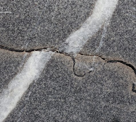
Stylolites or styolite (Greek: stylos, pillar; lithos, stone) are serrated surfaces within a rock mass at which mineral material has been removed by pressure dissolution, in a process that decreases the total volume of rock. Insoluble minerals, such as clays, pyrite and oxides, remain within the stylolites and make them visible. Sometimes host rocks contain no insoluble minerals, in which case stylolites can be recognized by change in texture of the rock.[1] They occur most commonly in homogeneous rocks,[2]carbonates, cherts, sandstones, but they can be found in certain igneous rocks and ice. Their size vary from microscopic contacts between two grains (microstylolites) to large structures up to 20 m in length and up to 10 m in amplitude in ice.[3] Stylolites usually form parallel to bedding, because of overburden pressure, but they can be oblique or even perpendicular to bedding, as a result of tectonic activity.[4][5]
Stylolites or styolite (Greek: stylos, pillar; lithos, stone) are serrated surfaces within a rock mass at which mineral material has been removed by pressure dissolution, in a process that decreases the total volume of rock. Insoluble minerals, such as clays, pyrite and oxides, remain within the stylolites and make them visible. Sometimes host rocks contain no insoluble minerals, in which case stylolites can be recognized by change in texture of the rock.[1] They occur most commonly in homogeneous rocks,[2]carbonates, cherts, sandstones, but they can be found in certain igneous rocks and ice. Their size vary from microscopic contacts between two grains (microstylolites) to large structures up to 20 m in length and up to 10 m in amplitude in ice.[3] Stylolites usually form parallel to bedding, because of overburden pressure, but they can be oblique or even perpendicular to bedding, as a result of tectonic activity.[4][5]
Structural geology is the study of the three-dimensional distribution of rock units with respect to their deformational histories. The primary goal of structural geology is to use measurements of present-day rock geometries to uncover information about the history of deformation (strain) in the rocks, and ultimately, to understand the stress field that resulted in the observed strain and geometries. This understanding of the dynamics of the stress field can be linked to important events in the geologic past; a common goal is to understand the structural evolution of a particular area with respect to regionally widespread patterns of rock deformation (e.g., mountain building, rifting) due to plate tectonics.
Structural geology is the study of the three-dimensional distribution of rock units with respect to their deformational histories. The primary goal of structural geology is to use measurements of present-day rock geometries to uncover information about the history of deformation (strain) in the rocks, and ultimately, to understand the stress field that resulted in the observed strain and geometries. This understanding of the dynamics of the stress field can be linked to important events in the geologic past; a common goal is to understand the structural evolution of a particular area with respect to regionally widespread patterns of rock deformation (e.g., mountain building, rifting) due to plate tectonics.
Structural geology is the study of the three-dimensional distribution of rock units with respect to their deformational histories. The primary goal of structural geology is to use measurements of present-day rock geometries to uncover information about the history of deformation (strain) in the rocks, and ultimately, to understand the stress field that resulted in the observed strain and geometries. This understanding of the dynamics of the stress field can be linked to important events in the geologic past; a common goal is to understand the structural evolution of a particular area with respect to regionally widespread patterns of rock deformation (e.g., mountain building, rifting) due to plate tectonics.
In optical mineralogy and petrography, a thin section (or petrographic thin section) is a laboratory preparation of a rock, mineral, soil, pottery, bones, or even metal sample for use with a polarizingpetrographic microscope, electron microscope and electron microprobe. A thin sliver of rock is cut from the sample with a diamond saw and ground optically flat. It is then mounted on a glass slide and then ground smooth using progressively finer abrasive grit until the sample is only 30 μm thick. The method involved using the Michel-Lévy interference colour chart. Typically quartz is used as the gauge to determine thickness as it is one of the most abundant minerals.
When placed between two polarizing filters set at right angles to each other, the optical properties of the minerals in the thin section alter the colour and intensity of the light as seen by the viewer. As different minerals have different optical properties, most rock forming minerals can be easily identified. Plagioclase for example can be seen in the photo on the right as a clear mineral with multiple parallel twinning planes. The large blue-green minerals are clinopyroxene with some exsolution of orthopyroxene.
Thin sections are prepared in order to investigate the optical properties of the minerals in the rock. This work is a part of petrology and helps to reveal the origin and evolution of the parent rock.
A photograph of a rock in thin section is often referred to as a photomicrograph.

 Turtledove Diffusers: Godox MF12 Wireless Flash (Set of 2)
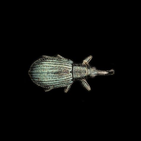 Imaging Single Specimens
 Turtledove Diffusers: Yongnuo YN-24EX TTL Macro Flash
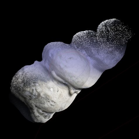 Internal Mold of a Turretellid Gastropod: Fossil