Showing 1–16 of 60 results

Copper

$19.00$400.00

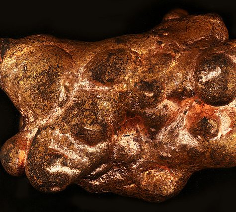
Copper is a chemical element with symbol Cu (from Latin: cuprum) and atomic number 29. It is a soft, malleable, and ductile metal with very high thermal and electrical conductivity. A freshly exposed surface of pure copper has a reddish-orange color. Copper is used as a conductor of heat and electricity, as a building material, and as a constituent of various metal alloys, such as sterling silver used in jewelry, cupronickel used to make marine hardware and coins, and constantan used in strain gauges and thermocouples for temperature measurement.

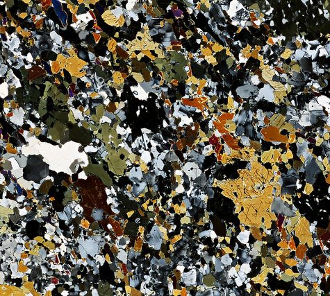
Copper is one of the few metals that occur in nature in directly usable metallic form (native metals) as opposed to needing extraction from an ore. This led to very early human use, from c. 8000 BC. It was the first metal to be smelted from its ore, c. 5000 BC, the first metal to be cast into a shape in a mold, c. 4000 BC and the first metal to be purposefully alloyed with another metal, tin, to create bronze, c. 3500 BC.

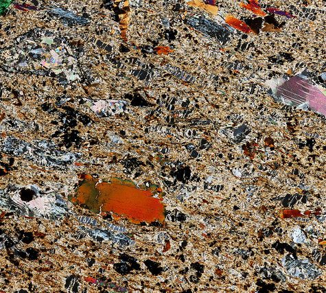
In the Roman era, copper was principally mined on Cyprus, the origin of the name of the metal, from aes сyprium (metal of Cyprus), later corrupted to сuprum, from which the words copper (English), cuivre (French), cobre (Spanish), Koper (Dutch) and Kupfer (German) are all derived. The commonly encountered compounds are copper(II) salts, which often impart blue or green colors to such minerals as azurite, malachite, and turquoise, and have been used widely and historically as pigments. Copper used in buildings, usually for roofing, oxidizes to form a green verdigris (or patina). Copper is sometimes used in decorative art, both in its elemental metal form and in compounds as pigments. Copper compounds are used as bacteriostatic agents, fungicides, and wood preservatives.

Copper is essential to all living organisms as a trace dietary mineral because it is a key constituent of the respiratory enzyme complex cytochrome c oxidase. In molluscs and crustaceans, copper is a constituent of the blood pigment hemocyanin, replaced by the iron-complexed hemoglobin in fish and other vertebrates. In humans, copper is found mainly in the liver, muscle, and bone. The adult body contains between 1.4 and 2.1 mg of copper per kilogram of body weight.

Wire Wrapped Pendant

$19.00$400.00

Wire wrapping is one of the oldest techniques for making handmade jewelry. This technique is done with some jewelry wire and findings similar to wire (like head-pins) to make components. Wire components are then connected to one another using mechanical techniques with no soldering or heating of the wire. Frequently, in this approach, a wire is bent into a loop or other decorative shape and then the wire is wrapped around itself to finish the wire component making that loop or decorative shape permanent. Because of this technique for wrapping wire around itself, this craft is called wire wrapping.

Reunion Island, 22,189, Old Glue disrupted optical properties 1x

$19.00$400.00

In optical mineralogy and petrography, a thin section (or petrographic thin section) is a laboratory preparation of a rock, mineral, soil, pottery, bones, or even metal sample for use with a polarizing petrographic microscope, electron microscope and electron microprobe. A thin sliver of rock is cut from the sample with a diamond saw and ground optically flat. It is then mounted on a glass slide and then ground smooth using progressively finer abrasive grit until the sample is only 30 μm thick. The method involved using the Michel-Lévy interference colour chart. Typically quartz is used as the gauge to determine thickness as it is one of the most abundant minerals.

When placed between two polarizing filters set at right angles to each other, the optical properties of the minerals in the thin section alter the colour and intensity of the light as seen by the viewer. As different minerals have different optical properties, most rock forming minerals can be easily identified. Plagioclase for example can be seen in the photo on the right as a clear mineral with multiple parallel twinning planes. The large blue-green minerals are clinopyroxene with some exsolution of orthopyroxene.

Thin sections are prepared in order to investigate the optical properties of the minerals in the rock. This work is a part of petrology and helps to reveal the origin and evolution of the parent rock.

A photograph of a rock in thin section is often referred to as a photomicrograph.

1x Hornblende-Plagioclase Gneiss 10,040 Adirondack Mountains

$19.00$400.00

In optical mineralogy and petrography, a thin section (or petrographic thin section) is a laboratory preparation of a rock, mineral, soil, pottery, bones, or even metal sample for use with a polarizing petrographic microscope, electron microscope and electron microprobe. A thin sliver of rock is cut from the sample with a diamond saw and ground optically flat. It is then mounted on a glass slide and then ground smooth using progressively finer abrasive grit until the sample is only 30 μm thick. The method involved using the Michel-Lévy interference colour chart. Typically quartz is used as the gauge to determine thickness as it is one of the most abundant minerals.

When placed between two polarizing filters set at right angles to each other, the optical properties of the minerals in the thin section alter the colour and intensity of the light as seen by the viewer. As different minerals have different optical properties, most rock forming minerals can be easily identified. Plagioclase for example can be seen in the photo on the right as a clear mineral with multiple parallel twinning planes. The large blue-green minerals are clinopyroxene with some exsolution of orthopyroxene.

Thin sections are prepared in order to investigate the optical properties of the minerals in the rock. This work is a part of petrology and helps to reveal the origin and evolution of the parent rock.

A photograph of a rock in thin section is often referred to as a photomicrograph.

5x Hornblende-Plagioclase Gneiss 10,040 Adirondack Mountains

$19.00$400.00

In optical mineralogy and petrography, a thin section (or petrographic thin section) is a laboratory preparation of a rock, mineral, soil, pottery, bones, or even metal sample for use with a polarizing petrographic microscope, electron microscope and electron microprobe. A thin sliver of rock is cut from the sample with a diamond saw and ground optically flat. It is then mounted on a glass slide and then ground smooth using progressively finer abrasive grit until the sample is only 30 μm thick. The method involved using the Michel-Lévy interference colour chart. Typically quartz is used as the gauge to determine thickness as it is one of the most abundant minerals.

When placed between two polarizing filters set at right angles to each other, the optical properties of the minerals in the thin section alter the colour and intensity of the light as seen by the viewer. As different minerals have different optical properties, most rock forming minerals can be easily identified. Plagioclase for example can be seen in the photo on the right as a clear mineral with multiple parallel twinning planes. The large blue-green minerals are clinopyroxene with some exsolution of orthopyroxene.

Thin sections are prepared in order to investigate the optical properties of the minerals in the rock. This work is a part of petrology and helps to reveal the origin and evolution of the parent rock.

A photograph of a rock in thin section is often referred to as a photomicrograph.

1x magnification, Hornblende-Plagioclase Gneiss 10,040 Adirondack Mountains

$19.00$400.00

In optical mineralogy and petrography, a thin section (or petrographic thin section) is a laboratory preparation of a rock, mineral, soil, pottery, bones, or even metal sample for use with a polarizing petrographic microscope, electron microscope and electron microprobe. A thin sliver of rock is cut from the sample with a diamond saw and ground optically flat. It is then mounted on a glass slide and then ground smooth using progressively finer abrasive grit until the sample is only 30 μm thick. The method involved using the Michel-Lévy interference colour chart. Typically quartz is used as the gauge to determine thickness as it is one of the most abundant minerals.

When placed between two polarizing filters set at right angles to each other, the optical properties of the minerals in the thin section alter the colour and intensity of the light as seen by the viewer. As different minerals have different optical properties, most rock forming minerals can be easily identified. Plagioclase for example can be seen in the photo on the right as a clear mineral with multiple parallel twinning planes. The large blue-green minerals are clinopyroxene with some exsolution of orthopyroxene.

Thin sections are prepared in order to investigate the optical properties of the minerals in the rock. This work is a part of petrology and helps to reveal the origin and evolution of the parent rock.

A photograph of a rock in thin section is often referred to as a photomicrograph.

5x magnification, Hornblende-Plagioclase Gneiss 10,040 Adirondack Mountains

$19.00$400.00

In optical mineralogy and petrography, a thin section (or petrographic thin section) is a laboratory preparation of a rock, mineral, soil, pottery, bones, or even metal sample for use with a polarizing petrographic microscope, electron microscope and electron microprobe. A thin sliver of rock is cut from the sample with a diamond saw and ground optically flat. It is then mounted on a glass slide and then ground smooth using progressively finer abrasive grit until the sample is only 30 μm thick. The method involved using the Michel-Lévy interference colour chart. Typically quartz is used as the gauge to determine thickness as it is one of the most abundant minerals.

When placed between two polarizing filters set at right angles to each other, the optical properties of the minerals in the thin section alter the colour and intensity of the light as seen by the viewer. As different minerals have different optical properties, most rock forming minerals can be easily identified. Plagioclase for example can be seen in the photo on the right as a clear mineral with multiple parallel twinning planes. The large blue-green minerals are clinopyroxene with some exsolution of orthopyroxene.

Thin sections are prepared in order to investigate the optical properties of the minerals in the rock. This work is a part of petrology and helps to reveal the origin and evolution of the parent rock.

A photograph of a rock in thin section is often referred to as a photomicrograph.

50x magnification, Hornblende-Plagioclase Gneiss 10,040 Adirondack Mountains

$19.00$400.00

In optical mineralogy and petrography, a thin section (or petrographic thin section) is a laboratory preparation of a rock, mineral, soil, pottery, bones, or even metal sample for use with a polarizing petrographic microscope, electron microscope and electron microprobe. A thin sliver of rock is cut from the sample with a diamond saw and ground optically flat. It is then mounted on a glass slide and then ground smooth using progressively finer abrasive grit until the sample is only 30 μm thick. The method involved using the Michel-Lévy interference colour chart. Typically quartz is used as the gauge to determine thickness as it is one of the most abundant minerals.

When placed between two polarizing filters set at right angles to each other, the optical properties of the minerals in the thin section alter the colour and intensity of the light as seen by the viewer. As different minerals have different optical properties, most rock forming minerals can be easily identified. Plagioclase for example can be seen in the photo on the right as a clear mineral with multiple parallel twinning planes. The large blue-green minerals are clinopyroxene with some exsolution of orthopyroxene.

Thin sections are prepared in order to investigate the optical properties of the minerals in the rock. This work is a part of petrology and helps to reveal the origin and evolution of the parent rock.

A photograph of a rock in thin section is often referred to as a photomicrograph.

Thin Section, 5x Polarized

$19.00$400.00

In optical mineralogy and petrography, a thin section (or petrographic thin section) is a laboratory preparation of a rock, mineral, soil, pottery, bones, or even metal sample for use with a polarizing petrographic microscope, electron microscope and electron microprobe. A thin sliver of rock is cut from the sample with a diamond saw and ground optically flat. It is then mounted on a glass slide and then ground smooth using progressively finer abrasive grit until the sample is only 30 μm thick. The method involved using the Michel-Lévy interference colour chart. Typically quartz is used as the gauge to determine thickness as it is one of the most abundant minerals.

When placed between two polarizing filters set at right angles to each other, the optical properties of the minerals in the thin section alter the colour and intensity of the light as seen by the viewer. As different minerals have different optical properties, most rock forming minerals can be easily identified. Plagioclase for example can be seen in the photo on the right as a clear mineral with multiple parallel twinning planes. The large blue-green minerals are clinopyroxene with some exsolution of orthopyroxene.

Thin sections are prepared in order to investigate the optical properties of the minerals in the rock. This work is a part of petrology and helps to reveal the origin and evolution of the parent rock.

A photograph of a rock in thin section is often referred to as a photomicrograph.

Thin Section, 1x

$19.00$400.00

In optical mineralogy and petrography, a thin section (or petrographic thin section) is a laboratory preparation of a rock, mineral, soil, pottery, bones, or even metal sample for use with a polarizing petrographic microscope, electron microscope and electron microprobe. A thin sliver of rock is cut from the sample with a diamond saw and ground optically flat. It is then mounted on a glass slide and then ground smooth using progressively finer abrasive grit until the sample is only 30 μm thick. The method involved using the Michel-Lévy interference colour chart. Typically quartz is used as the gauge to determine thickness as it is one of the most abundant minerals.

When placed between two polarizing filters set at right angles to each other, the optical properties of the minerals in the thin section alter the colour and intensity of the light as seen by the viewer. As different minerals have different optical properties, most rock forming minerals can be easily identified. Plagioclase for example can be seen in the photo on the right as a clear mineral with multiple parallel twinning planes. The large blue-green minerals are clinopyroxene with some exsolution of orthopyroxene.

Thin sections are prepared in order to investigate the optical properties of the minerals in the rock. This work is a part of petrology and helps to reveal the origin and evolution of the parent rock.

A photograph of a rock in thin section is often referred to as a photomicrograph.

Thin Section, 1x Polarized

$19.00$400.00

In optical mineralogy and petrography, a thin section (or petrographic thin section) is a laboratory preparation of a rock, mineral, soil, pottery, bones, or even metal sample for use with a polarizing petrographic microscope, electron microscope and electron microprobe. A thin sliver of rock is cut from the sample with a diamond saw and ground optically flat. It is then mounted on a glass slide and then ground smooth using progressively finer abrasive grit until the sample is only 30 μm thick. The method involved using the Michel-Lévy interference colour chart. Typically quartz is used as the gauge to determine thickness as it is one of the most abundant minerals.

When placed between two polarizing filters set at right angles to each other, the optical properties of the minerals in the thin section alter the colour and intensity of the light as seen by the viewer. As different minerals have different optical properties, most rock forming minerals can be easily identified. Plagioclase for example can be seen in the photo on the right as a clear mineral with multiple parallel twinning planes. The large blue-green minerals are clinopyroxene with some exsolution of orthopyroxene.

Thin sections are prepared in order to investigate the optical properties of the minerals in the rock. This work is a part of petrology and helps to reveal the origin and evolution of the parent rock.

A photograph of a rock in thin section is often referred to as a photomicrograph.

Thin Section, 5x Polarized

$19.00$400.00

In optical mineralogy and petrography, a thin section (or petrographic thin section) is a laboratory preparation of a rock, mineral, soil, pottery, bones, or even metal sample for use with a polarizing petrographic microscope, electron microscope and electron microprobe. A thin sliver of rock is cut from the sample with a diamond saw and ground optically flat. It is then mounted on a glass slide and then ground smooth using progressively finer abrasive grit until the sample is only 30 μm thick. The method involved using the Michel-Lévy interference colour chart. Typically quartz is used as the gauge to determine thickness as it is one of the most abundant minerals.

When placed between two polarizing filters set at right angles to each other, the optical properties of the minerals in the thin section alter the colour and intensity of the light as seen by the viewer. As different minerals have different optical properties, most rock forming minerals can be easily identified. Plagioclase for example can be seen in the photo on the right as a clear mineral with multiple parallel twinning planes. The large blue-green minerals are clinopyroxene with some exsolution of orthopyroxene.

Thin sections are prepared in order to investigate the optical properties of the minerals in the rock. This work is a part of petrology and helps to reveal the origin and evolution of the parent rock.

A photograph of a rock in thin section is often referred to as a photomicrograph.

Thin Section, 1x Polarized

$19.00$400.00

In optical mineralogy and petrography, a thin section (or petrographic thin section) is a laboratory preparation of a rock, mineral, soil, pottery, bones, or even metal sample for use with a polarizing petrographic microscope, electron microscope and electron microprobe. A thin sliver of rock is cut from the sample with a diamond saw and ground optically flat. It is then mounted on a glass slide and then ground smooth using progressively finer abrasive grit until the sample is only 30 μm thick. The method involved using the Michel-Lévy interference colour chart. Typically quartz is used as the gauge to determine thickness as it is one of the most abundant minerals.

When placed between two polarizing filters set at right angles to each other, the optical properties of the minerals in the thin section alter the colour and intensity of the light as seen by the viewer. As different minerals have different optical properties, most rock forming minerals can be easily identified. Plagioclase for example can be seen in the photo on the right as a clear mineral with multiple parallel twinning planes. The large blue-green minerals are clinopyroxene with some exsolution of orthopyroxene.

Thin sections are prepared in order to investigate the optical properties of the minerals in the rock. This work is a part of petrology and helps to reveal the origin and evolution of the parent rock.

A photograph of a rock in thin section is often referred to as a photomicrograph.