This pamphlets below help describe the imaging capabilities offered by the Macropod Pro imaging system. The “Macropod” can be used to photograph everything from outcrops to mineral and/or fossil specimens (even thin sections), almost to the level of a scanning electron microscope (SEM). The unique thing is that the host company, Macroscopic Solutions, provides an all-in-one portable professional imaging set up (backpack, camera, numerous lenses, stands, image stacking software, etc…) to conduct field- and lab-based professional 3D imagery, making it a fantastic tool for education and outreach.
To get a sense of what I’m talking about, you can look at the following links included below:
Colleagues that have already purchased one of the Macropods for his or her department are heavily used by faculty and graduate students for everything from research-based analysis to classwork on cores, specimens, and outcrop-based images.
The CUIC at Cornell University recently published a post about how effective their system has been (see number 2).

Macropod Pro captured all color images, which are higher in resolution than the B&W SEM imagery.

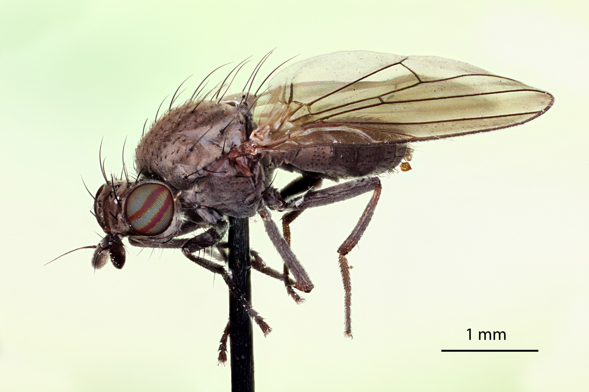
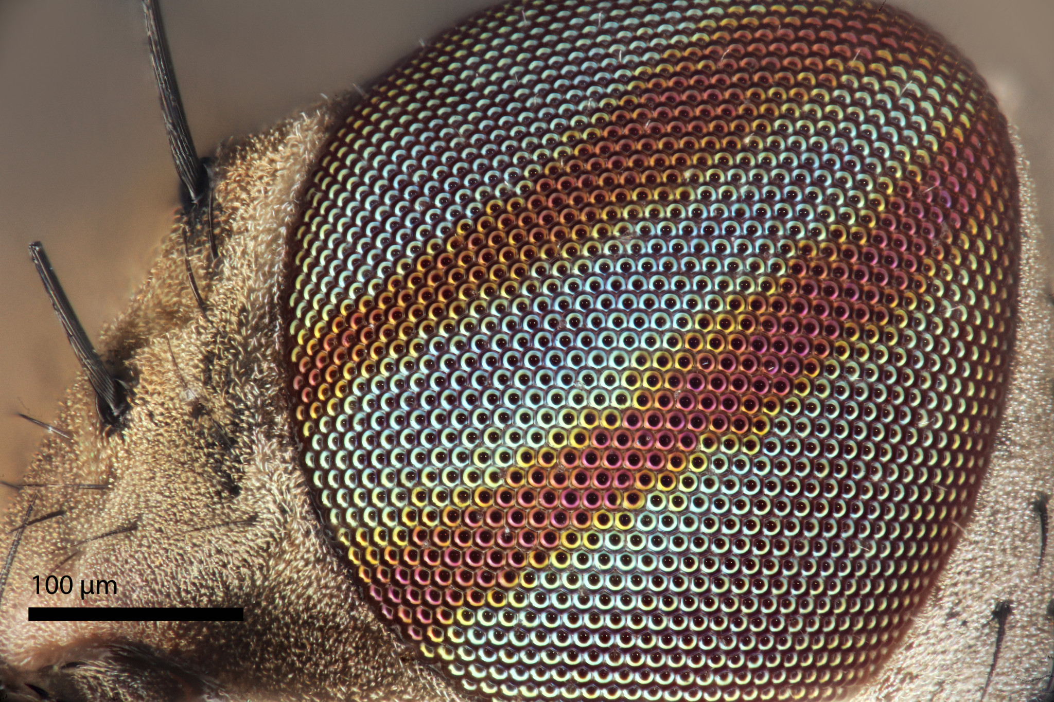
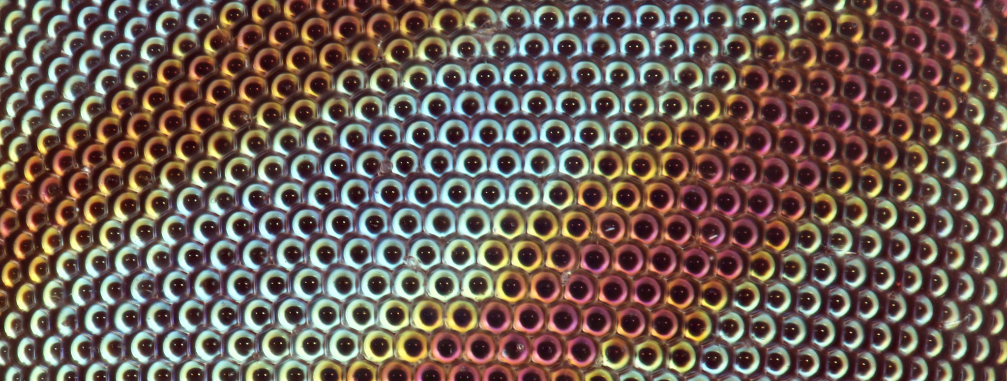
Sapromyza brachysoma, Fly, Coventry, CT
Early Stage Mouse Embryo (Blood Cells) Shot at 5x with Fluorescence

20x Pine Needles imaged using Fluorescence

10x Pine Needles imaged using Fluorescence